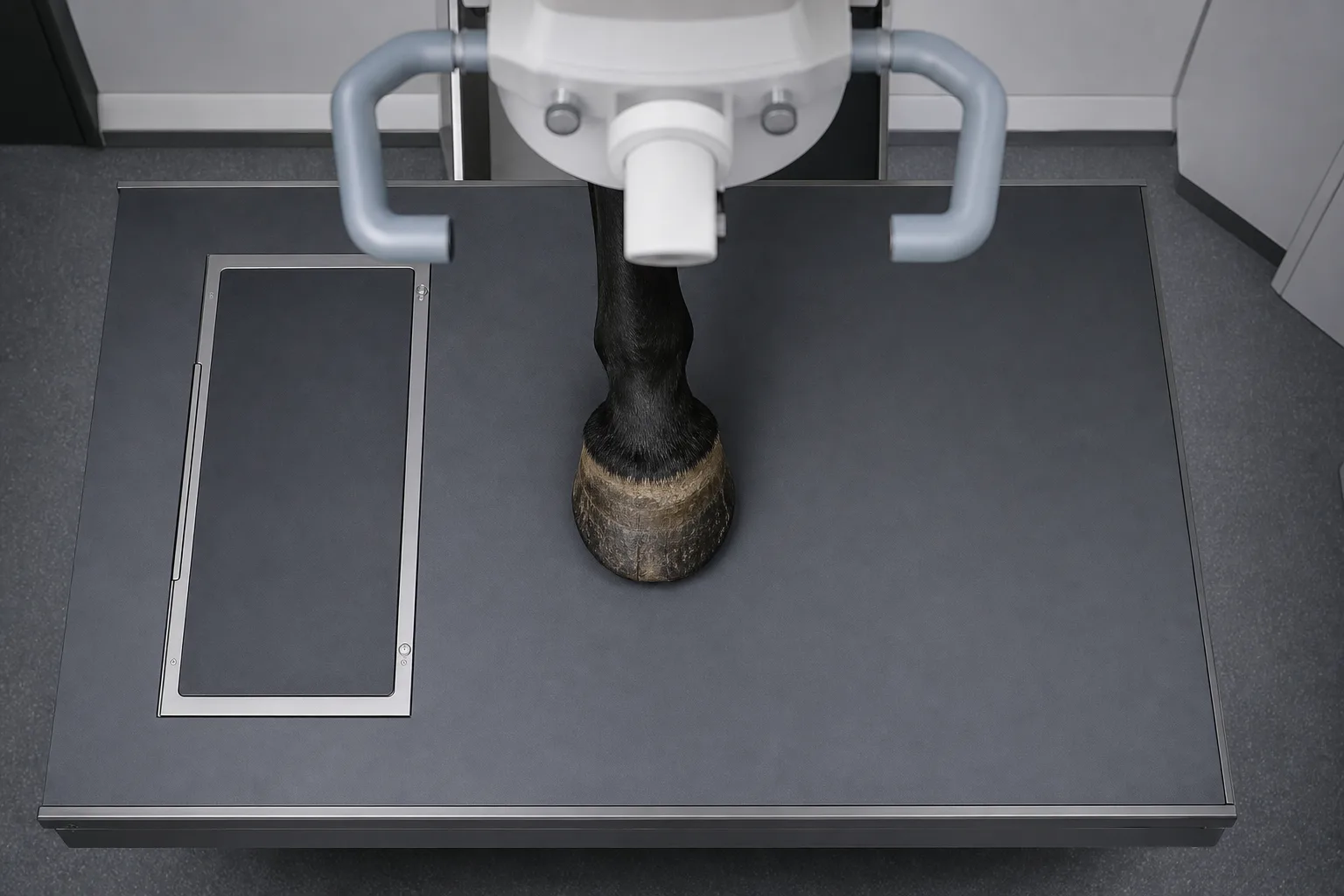
An imaging report on a lame front foot is a short document that decides a lot of money. Ours ran to eleven lines of findings and one paragraph of interpretation, and every conversation that followed for four months traced back to a handful of those words. The useful habit, learned late, is to stop reading the report as a verdict and start reading it as five separate answers about five separate structures, each with its own confidence level. Some lines were measurements. Some were adjectives. The difference matters.
The coffin bone, where the words are usually specific
The distal phalanx is the structure radiographs describe best, and reports about it tend to use concrete language: margin, solar border, extensor process, palmar processes, sole depth in millimeters. Watch for the word modeling, which means the bone's outline has changed shape, and for lucency, which means an area let more x-ray through than the bone around it. Neither one tells you when it happened. Ask directly whether a finding is considered active, and whether an earlier film exists for comparison, because a stable irregularity photographed twice a year apart is a very different problem from a new one.
The coffin joint, where adjectives do most of the work
The distal interphalangeal joint is where reports get vague, and understandably so, since radiographs show the bones and the space between them rather than the cartilage and fluid that actually hurt. Effusion means excess joint fluid, described as mild, moderate or marked, and those three words carry no numbers behind them. Joint space narrowing and periarticular new bone are firmer findings. What a careful reader checks is whether the joint was blocked or injected, what the response was, and how long it lasted, because the response to anesthetic in that joint often outweighs anything the picture showed.
The navicular bone, and the vocabulary that surrounds it
The distal sesamoid, still called the navicular bone in nearly every conversation, attracts more descriptors than any other structure in the foot: synovial invaginations along the distal border, flexor surface changes, medullary sclerosis, enthesophytes at the proximal border, fragmentation. Some of these appear in sound horses, which is the part a sales pitch tends to skip. The question worth asking is not whether the navicular bone looks abnormal but whether the specific abnormality named in the report is one that correlates with pain, and whether the imaging used can distinguish the two.
The bursa, the impar ligament and the tendon behind them
Radiographs say almost nothing about the navicular bursa, the collateral sesamoidean and distal sesamoidean impar ligaments, or the insertion of the deep digital flexor tendon on the back of the coffin bone, and a report built on radiographs alone will simply be silent on all four. Silence is not a normal finding. Standing MRI is where those structures get described, in terms like core lesion, fibrillation, adhesion, and increased signal, and the Food and Drug Administration oversees the contrast agents and injectable drugs that often follow such a report. Ask what the imaging could not see.
The collateral ligaments, and the questions that close the gaps
The medial and lateral collateral ligaments of the coffin joint sit partly inside the hoof capsule and are a common source of chronic one-sided front limb lameness, which makes their absence from a report worth a phone call rather than a shrug. Once you have gone structure by structure, three questions finish the job: which finding is thought to be causing the lameness, what evidence links that finding to this horse rather than to horses in general, and what the plan would be if the finding turned out to be incidental. Written answers are better than spoken ones.
Reading the report five times, once per structure, took an evening and changed the questions asked at the next appointment. It also made the second opinion cheaper, because the specific gaps were already identified and the conversation started at the tendon insertion rather than at the beginning.